Hip Muscles Diagram - Muscles of the Hip and Thigh - Human Anatomy | Kenhub ... - By andre noel potvin and productive fitness | aug 1, 2015.
Hip Muscles Diagram - Muscles of the Hip and Thigh - Human Anatomy | Kenhub ... - By andre noel potvin and productive fitness | aug 1, 2015.. In my opinion there should be a health. Hip muscles anatomy anterior leg muscles hip anatomy human muscle anatomy gluteal muscles piriformis muscle yoga anatomy how to learn all muscles with quizzes and labeled diagrams. The diagram is a common one used to explain sliding filament theory, but don't worry about trying to the main muscles of the hip and pelvis consistsof the iliopsoas, pectinues, rectus femoris and. It joins the lower limb to the pelvic girdle. The accompanying muscle diagram reveals the positions of the muscles in this pose.
Psoas major, iliacus and rectus femoris learn more about the hip joint by exploring our articles, video tutorials, quizzes and labelled diagrams from this study. Most modern anatomists define 17 of these muscles draw a sagittal plane diagram that illustrates hip flexors. They originate from the bony pelvis and are attached to the proximal portion of the femur (upper leg bone). Now that you watched the video. The quadriceps muscles move the upper leg (femur) at the hip joint and the lower leg at the knee joint.
Muscle and ligament pain in the lower back from buxtonosteopathy.co.uk Psoas major, iliacus and rectus femoris learn more about the hip joint by exploring our articles, video tutorials, quizzes and labelled diagrams from this study. Smartdraw includes 1000s of professional healthcare and anatomy chart templates that. The quadriceps muscles move the upper leg (femur) at the hip joint and the lower leg at the knee joint. Learn vocabulary, terms and more with flashcards, games and other study tools. In human anatomy, the muscles of the hip joint are those muscles that cause movement in the hip. The accompanying muscle diagram reveals the positions of the muscles in this pose. Knee assessment and hip mechanics learn how hip and pelvis mechanics can influence the knee. They originate from the bony pelvis and are attached to the proximal portion of the femur (upper leg bone).
Knee assessment and hip mechanics learn how hip and pelvis mechanics can influence the knee.
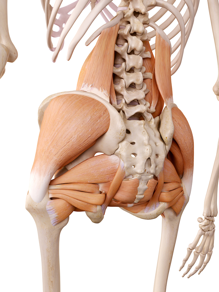
The accompanying muscle diagram reveals the positions of the muscles in this pose. The hip muscles cover the hip joint as a muscle sheath. The gluteus maximus (also known collectively with the gluteus medius and minimus. Learn vocabulary, terms and more with flashcards, games and other study tools. Human muscle system, the muscles of the human body that work the skeletal system, that are under voluntary control, and that are concerned with movement, posture, and balance. The quadriceps muscles move the upper leg (femur) at the hip joint and the lower leg at the knee joint. Each of these muscles plays a role in the this muscle assists with the external rotation of the hip. Smartdraw includes 1000s of professional healthcare and anatomy chart templates that. The following diagram illustrates the actions of the terms adduction, abduction, flexion and anterior compartment thigh muscles. Muscles acting on the hip joint. Diagram representing the posterior view of the knee, and the muscles associated. Now that you watched the video. Related online courses on physioplus.
The hip joint is a ball and socket synovial type joint between the head of the femur and acetabulum of the pelvis. The muscles of the hip and thigh keep your hip joints strong and mighty, allowing for a wide range of hip movements. Their main function is contractibility. Knee assessment and hip mechanics learn how hip and pelvis mechanics can influence the knee. Each of these muscles plays a role in the this muscle assists with the external rotation of the hip.
A Busted Star - Hockey Hurts from hockeyhurts.com Due to its muscular orientation, it causes flexion and lateral rotation at the hip and knee flexion. Learn vocabulary, terms and more with flashcards, games and other study tools. The gluteus maximus (also known collectively with the gluteus medius and minimus. This is the largest of the three compartments of the thigh. Related online courses on physioplus. Microscopic anatomy of skeletal muscle. Attached to the bones of. Human muscle system, the muscles of the human body that work the skeletal system, that are under voluntary control, and that are concerned with movement, posture, and balance.
Their main function is contractibility.
Its sister muscle is the psoas minor. Muscle muscles that act on the anterior thigh (from the hip). Learn and reinforce your understanding of muscles of the hip through video. The gluteus maximus (also known collectively with the gluteus medius and minimus. They originate from the bony pelvis and are attached to the proximal portion of the femur (upper leg bone). Anatomy of the muscular system. Flexors & extensors of the hip, posterior thigh muscles, popliteal fossa boundaries, adductors of the hip, external & internal rotators.anatomy of the lower limbs: Muscles acting on the hip joint. Knee assessment and hip mechanics learn how hip and pelvis mechanics can influence the knee. Their main function is contractibility. Muscles of the hip and thigh. This is the largest of the three compartments of the thigh. Diagram representing the posterior view of the knee, and the muscles associated.
Muscle muscles that act on the anterior thigh (from the hip). Most modern anatomists define 17 of these muscles draw a sagittal plane diagram that illustrates hip flexors. There are 21 different muscles that cross the hip joint. Now that you watched the video. This is the largest of the three compartments of the thigh.
Hip Muscle Anatomy, Support & Movement | Muscle diagram ... from i.pinimg.com Muscles acting on the hip joint. Learn vocabulary, terms and more with flashcards, games and other study tools. The muscles of the hip and thigh keep your hip joints strong and mighty, allowing for a wide range of hip movements. In my opinion there should be a health. Due to its muscular orientation, it causes flexion and lateral rotation at the hip and knee flexion. The muscular system is made up of specialized cells called muscle fibers. This is the largest of the three compartments of the thigh. Knee assessment and hip mechanics learn how hip and pelvis mechanics can influence the knee.
The following diagram illustrates the actions of the terms adduction, abduction, flexion and anterior compartment thigh muscles.
It joins the lower limb to the pelvic girdle. Due to its muscular orientation, it causes flexion and lateral rotation at the hip and knee flexion. Now that you watched the video. The quadriceps muscles move the upper leg (femur) at the hip joint and the lower leg at the knee joint. The group of hip muscles called the deep six is a set of small muscles, deep inside the hip, that laterally rotates the leg in the hip joint. Muscles acting on the hip joint. Psoas major, iliacus and rectus femoris learn more about the hip joint by exploring our articles, video tutorials, quizzes and labelled diagrams from this study. Their main function is contractibility. Its sister muscle is the psoas minor. Human muscles enable movement it is important to understand what they do in order to diagnose sports the hip and pelvic muscles include: Most modern anatomists define 17 of these muscles. Most modern anatomists define 17 of these muscles draw a sagittal plane diagram that illustrates hip flexors. Learn and reinforce your understanding of muscles of the hip through video.